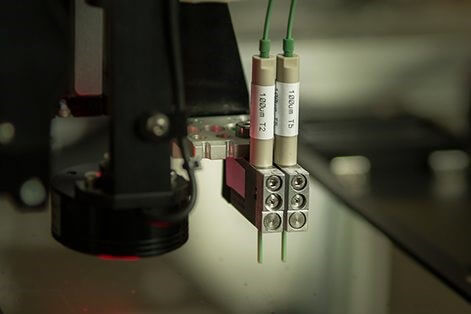
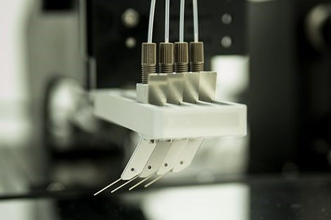
LAMINATION/CUTTING OF LFA STRIPS:

LM5000

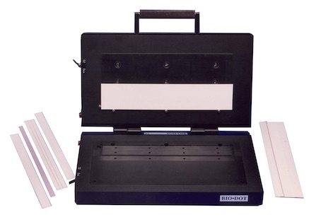
The LM5000 Clamshell assembles a lateral flow assay comprised of multiple materials onto an adhesive backing. It contains top and bottom vacuum nests to hold strip materials in place and is operated manually. Standard and customized nest inserts are available and are easily interchangeable so that multiple assay designs can be laminated.

LM9000

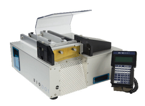
The LM9000 is a continuous laminator which provides continuous lamination of materials onto a plastic support backing with adhesive. This would be used for a assembling rapid diagnostic test strips.

CM5000

The CM5000 guillotine cutter is also available to provide high quality precision cuts. The widths and cuts can be programmed through a handheld terminal.

DISPENSE PLATFORMS, continued:

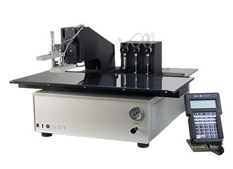
The XYZ series systems are a powerful production tool for rapid test development and manufacturing with a larger working deck than the smaller XYZ3060 platform.

XYZ3210

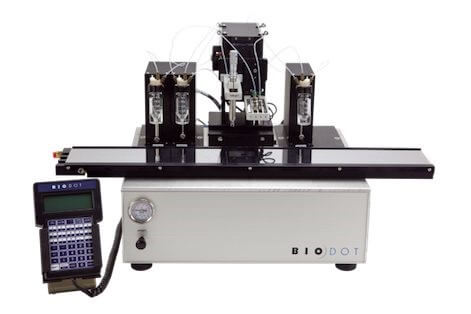
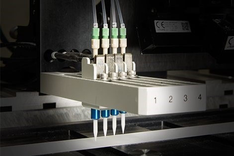
The XYZ3210 is a flexible XYZ motion/dispense system, with a motored Z-axis and fully programmable motion and dispense parameters. The XYZ3210 is designed for simultaneous dispensing or individually dispensing lines and dots. This platform can incorporate 4 AirJet HR, 8 BioJet HR, 8 FrontLine HR, or 8 PolyDrop dispensers. The researcher can choose the type and number of dispensers systems for their application needs.

XYZ3060

The XYZ3060 system is similar to the XYZ321- system, but with a smaller working deck.

BioDot also provides a variety of automated camera vision solutions in order to increase reproducibility in manufacturing as well as reliability in part inspection. Automated inspection process can lead to a higher quality of product and increased yield. All of these LFA dispensing systems to create your own design and applications can be seen on BioDot’s web site.: https://www.biodot.com/lateral-flow/

Continue to next article: Manufacturing of Lateral Flow Assays (Part 6)

BioDot provides a number of reagent dispensing platforms that are designed for a combination of R&D and production activities. In both cases, the same dispensing technology is used which allows for R&D validated processes to be efficiently transferred to the manufacturing environment.

IMPREGNATION:
Bulk impregnation of sheets in baths or in-line with the RR120 is available. The RR120 has an impregnation module for dipping web materials followed by drying. However, neither of these options are recommended due to the basic non-homgenous nature of these materials which leads to the variations of dried reagent concentrations. This problem is eliminated by the use of the previously described dispensing reagent systems (BioJet HR, AirJet Ultra, AirJet HR, etc). For continuous dispensing, a tandem pump configuration is used for both the FrontLine and BioJet dispensing. BioDot has international patents (US, China, Europe) for the BioJet HR™, BioJet Ultra™, AirJet, as well as the used of solenoid and aerosol dispensed heads combined with the use of syringe pumps to achieve quantitative dispensing. BioDot’s additional patents include the use of the tandem pump configuration, which eliminates delays and line distortions due to syringe refilling cycles.


DISPENSE PLATFORMS:

ZX1010 Platform

The ZX1010 is a flexible dispensing system with a programmable X-axis, manually adjusted Y-axis positioning, and a pneumatic Z-axis. Dispensed volumes are independently programmed via the terminal.

The ZX1010 platform can be configured with a maximum of 8 dispensers (max 4 AirJets). You can choose the type and number of dispensers to meet your application needs and includes the choices of the µAirJet, AirJet HR, BioJet HR, Frontline HR, and PolyDrop dispensers.

Continue to next article: Manufacturing of Lateral Flow Assays (Part 5)

DISPENSING AND IMPREGNATION PRODUCTS, continued:

AirJet HR™

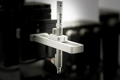
The AirJet HR™ is a nanoliter aerosol dispenser. This BioDot system uses pressurized air to atomize fluid passing through the dispensing nozzle for non-contact, quantitative aerosol dispensing. This creates a quantitative spray format, in which a dot or line can be quantitatively generated on a continuous basis. BioDot’s proprietary technology couples the dispensing nozzle with a high resolution syringe pump to meter exact amounts of reagents. This process produces a precise and easy to use method for dispensing micoliter quantities of fluids.

BioDot’s proprietary technology couples the dispensing nozzle with a high resolution syringe pump to meter exact amounts of reagents. This proces produces a precise and easy to use method for dispensing micoliter quantities of fluids.

µAirJet™

The µAirJet™ is another aerosol type dispenser which uses pressurized air to atomize fluid passing through the nozzle. This nozzle is too, coupled with a syringe pump which creates a spray format. The straighforward design makes µAirJet easy to use, clean, and maintain.

Continue to next article: Manufacturing of Lateral Flow Assays (Part 4)

DISPENSING AND IMPREGNATION PRODUCTS, continued:
The next several systems are non-contact, which is the preferred system with LFA diagnostic assay applications. All of these are offered by BioDot.

BioJet HR™

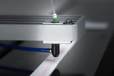
The BioJet HR™ is a proprietary, qualitative, non-contact technology which couples the BioJet “Drop-on-Demand” valve with a high-resolution syringe pump. The BioJet HR™ meters precise amounts of reagent, incorporating the benefits of non-contact dispensing and the ability to program exact drop volumes (nanoliter to microliter).

BioJet Ultra™

The BioJet UltraTM is a non-contact system capable of handling picoliter to nanoliter liquid handling and spotting solution with drop-on-demand capability. The dispense platform can produce a single dynamic drop volume range of 100pL to 1.0nL. It is compatible with a wide range of reagent types, including aqueous, organic, and cells. The platform can be optimized to work with a high dynamic range of viscosities without additives (up to 400 cP).

Continue to next article: Manufacturing of Lateral Flow Assays (Part 3)

INTRODUCTION:
There are many companies who provide equipment for making diagnostic LFAs. The LFA device is a test strip inside a plastic housing. This housing can have two or more ports for sample analysis. The strips have five components which include: 1) plastic backing with adhesive and release liner, 2) sample pad, 3) conjugate pad, 4) nitrocellulose pad, and 5) cover layers over the composite device. The materials are supplied in a roll, sheet or strip formats. The membrane is already converted for lamination onto the plastic backing. Other materials are converted to the correct widths for processing and lamination before, or after, reagents are process. In the next several short notes, I will present how one company, BioDot, makes their LFA devices, using information and product photos taken from their website https://www.biodot.com/lateral-flow/.

MEMBRANE:
Dispensing test and control lines onto the membrane can be done before or after lamination. Blocking reagents can be dispensed in parallel with the test and control, or afterwards. The typical dispensing onto the membrane is in a strip format using XY tables with bare membrane before lamination or maters strips where the membrane has been laminated onto the plastic backing. The strip length is typically 300-500 mm. The blocking reagent can also be dispensed using a third dispense channel in parallel with the test and control lines. This dispensing can also be done on a reel-to-reel system using a roll format where the membrane is unbacked or pre-laminated to the backing material. Line dispensing can be done with a contact or non-contact dispensing system. The non-contact is preferable, as contact dispensing leaves a small indentation on the membrane surface that degrades the quality of the dispensed line.

CONJUGATE AND SAMPLE PADS:
Two pads are typically processed using impregnated sheet stock, followed by drying, cutting to width, and lamination to the backing card. Web materials can be impregnated with an in-line reel-reel machine. The reagent is very accurately dispensed along the lamination direction so that at each cut the lateral flow strip will have the same volume or dried reagent.


DISPENSING AND IMPREGNATION PRODUCTS:
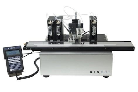
BioDot provides a number of dispensing and impregnation options for processing lateral flow test strips formats that can be integrated into different platforms. One such system is their Frontline HFTM  platform, shown below.

The Frontline HFTM system is ideal for printing lines on membranes and other substrates for rapid test, immunoblots and biochips. However, since the dispensing tip rides on the substrate surface, it leaves a small indentation on the membrane surface that degrades the quality of the dispensed line. For this reason, it is not preferable for use with a lateral flow system.

Continue to next article: Manufacturing of Lateral Flow Assays (Part 2)

INTRODUCTION:
Relatively few multiplex lateral flow immunoassays (LFIAs) are available today for detecting viral pathogens, primarily due to technical and manufacturing hurdles. However, several of these assays are being developed. The detection of antibodies against more than one agent is probably the easiest type of multiplex assay to manufacture. The key components of such assays are to have specific antigen targets immobilized on a membrane strip which can be detected by the antibodies in a patient’s sera. However, the demand for such tests need to by evaluated as to their market need and whether it is commercially feasible to offer multiplexed assays, rather than monospecific assays for each target.

DISCUSSION:
Anfossi et al1Anfossi et al., 2018. Multiplex Lateral Flow Immunoassay: An Overview of Strategies towards High-throughput Point-of-Need Testing. Biosensors 9 (2), 1-14., give an excellent synopsis of developing multiplex assays using either spatially separated areas (test lines) on the same strip, or a platform which uses multiple strips for each analyte to be detected. A wide variety of multiplex LFIA have been developed to detect antibiotics, drugs, chemicals, poisons and toxins; Song et al.2Song et al., 2015. Multiplex Lateral Flow Immunoassay for Mycotoxin Determination. Anal. Chem. 86, 4995-5001., Peng et al.3Peng et al., 2016. Multiplex lateral flow immunoassay for five antibiotics detection based on gold nanoparticle aggregations. RSC Adv. 6, 7798-7805., and Xing et al44) Xing et al., Ultrasensitive immunochromatographic assay for the simultaneous detection of five chemicals in drinking water. Biosens. Bioelectron. 66, 445-453.. Far fewer have been developed to detect viral pathogens. An example for one of the multiplex LFIA for a viruses is the two-color assay developed by Lee et al.5Lee et al., 2016. Two-color Lateral Flow Assay for Muliplex Detection of Causative Agents Behind Acute Febrile Illness. Anal. Chem. 88, 8359-8363.. Their goal was to use a patient’s serum antibodies to detect 2 different pathogens (dengue and Chikungunya viruses) on the same test strip which also detected both IgM (an indicator of a recent infection) and IgG (an indicator for a past infection) against each virus. This novel assay is actually a 4-plex system, detecting both IgG and IgM for both dengue and Chikungunya viruses. To accomplish this, they used colored latex beads to detect each antibody (blue latex for anti-IgG and red latex for anti-IgM). Lee describes this assay as: the sample pad for accepting the sample, the conjugate pad for storing the detection probes, the nitrocellulose membrane for immobilizing the test and control region reagents, and the absorbent pad for collecting the waste mixtures. The samples (blood/plasma/serum) and chase buffer are added to the sample pad. Once wetted, the blue and red detection probes which have been dry-stored on the conjugate pad are released and become free to interact with the sample IgGs and IgMs which have bound to their respective recombinant viral protein targets in the test line. Further downstream on the strip, secondary antibodies (i.e. anti-goat IgG) capture both blue anti-IgG and red anti-IgM detection probes based on their host species (i.e. goat) and develop mixed colors to confirm that the test has completed successfully.

CONCLUSION:
The paper by Lee et al.6Lee et al., 2016. Two-color Lateral Flow Assay for Muliplex Detection of Causative Agents Behind Acute Febrile Illness. Anal. Chem. 88, 8359-8363., was the first demonstration of the ability to detect both IgG and IgM for more than one pathogen on a single strip. They showed that by observing the color and location of the CHIKV and DENV test regions, their assay could indicate the presence of anti-CHIKV IgG/IgM and anti-DENV IgG/IgM. This approach should be applicable for detecting other multiple pathogen recombinant proteins on a single strip. However, it is mentioned that, as the number of test regions increases there may be a need for a simple analyzer detection system to accommodate users who may have color-perception difficulties.

INTRODUCTION:
Lateral flow immunoassays (LFIAs) are an important component in point-of-care patient diagnostics. More LFIAs are being developed every year, driven by the need of rapid, low-cost information in a patient or hospital setting. Presented in this note will be the overall advantages and disadvantages to LFIAs, as well as new research to improve the lateral flow assay (LFA) technology.

DISCUSSION:
LFAs can be classified into two types of basic assays. One is nucleic acid lateral flow assay (NALFA) and the other is the lateral flow immunoassay (LFIA). The LFIA has several components which need to be carefully assembled and tested in assay development. All of these are described in detail by Koczula and Gallotta1Koczula, K.M., and Andrea Gallota. 2016. Lateral Flow assays. Essays in Biochemistry. 60, 111-120., and include 1) the antibody, which needs to be carefully designed and highly purified, 2) the nanoparticle label, which needs to be stable, easily reproducible, efficient, commercially available and easy to scale up the conjugation procedure, 3) the LFIA membrane strip, which is most often nitrocellulose, but which must have a uniform pore size and good binding characteristics to the proteins in the assay, 4) the sample pad which has the buffer salts proteins, surfactants and other liquids to control the flow rate across the strip, 5) the conjugate pad, of which the main role is to hold the detector particles and keep them stable, and 6) the absorbent pad for wicking the fluid through the membrane and to collect the processed liquid. One of the most important benefits of an LFIA is that it is usually a one-step assay which requires no special skills or instrumentation to achieve the result(s). The fact that these types of assays are qualitative, yes/no, leads to its simple determination. These tests can be done at the point-of-care, or even in the patient’s home (the self-pregnancy test which detects the hCG hormone is probably the most widely known LFA on the market). In the case of LFIAs for pathogens, the assay targets can be pathogen specific proteins, antibodies, or nucleic acids. These assays usually have a long shelf life and do not require refrigeration or freezer storage of the assay reagents. Finally, the samples do not normally need to be pre-treated before applying to the LFIA. However, there are several draw-backs with the LFIA technology. Applying the wrong amount of sample onto the LFIA can test strip can alter the reliability of the test results. Antibody preparation is a critical step for the LFIA. Sometimes the nature of the sample can alter the assay results, or the time needed for the assay to “develop”.  The nature of the sample can also alter the capillary action, or spread, of the target molecule on the test strip. In cases, such as this, pre-treatment of the sample may be needed. And finally, although the nature of the LFIA leads to low costs for the end user, there can be very large development costs in the design/development of the assays by the manufacturer.

CONCLUSION:
Lateral flow assays, or more specifically, LFIAs have a wide range of application for detecting biological pathogens in a clinical and non-clinical setting. What the LFIA assays lack in sensitivity is made up for by its low cost, rapid results and simplicity of use. Although most LFIAs (or LFAs) are monospecific, more assays are being developed in a multiplex format for detecting more than one target from a specific pathogen, or for detecting more than one pathogen. New strategies are being devised to increase the sensitivity by enhancing the signal using colloidal gold nanoparticles, or using gold nanoparticles in combination with horseradish peroxidase, which results in an amplification of the signal2Parolo, C., de la Escosura-Muniz, A. and Merkoci, A. 2013. Enhanced lateral flow immunoassay using gold nanoparticles loaded with enzymes. Biosens. Bioelectron. 40, 412-416.. A combination of detecting both antigens and antibodies by using two conjugate pads for the simultaneous detection of two proteins has also been described3Zhu, J., Zou, N., Mao, H., Wang, P., Zhu, D., Ji, H. et al., 2013. Evaluation of a modified lateral flow immunoassay for detection of high-sensitivity cardiac troponin I and myoglobin. Biosens. Bioelectron. 42, 522-525..

INTRODUCTION:
In the last several years there has been an increasing demand for the development of infectious disease diagnostics which maintains test accuracy while reducing the time needed for detection at the point-of-care (POC) for the patient. Reducing the cost per assay as well as the equipment and end-user technical knowledge has been the driving force between the development of the lateral flow assay. This is primarily a qualitative colorimetric assay which can detect pathogen related proteins, nucleic acids, or antibodies.  Here is a brief summary of several recent reviews for LFAs.

DISCUSSION:
Most conventional clinical diagnostics offers high specificity for the detection of specific pathogens. This specificity comes at the high cost of time, equipment and technical expertise to run these tests, not to mention the need to adhere to CLIA guidelines and testing regimens for a clinically approved assay. The LFA can overcome all of these obstacles. Basically, the LFA is a membrane (nitrocellulose or glass fiber substrate) which contains absorbed reagents which react to the applied sample in order to detect the target molecule (usually a pathogen related protein or specific antibody). However, other pathogen specific molecules can be applied to the LFA, such as aptamers (artificial nucleic acids which can selectively bind to target analytes),  molecular beacons (a special DNA hairpin structure with an attached fluorophore which does not produce fluorescence in the absence of an analyte, but when a complementary DNA sequence (target nucleic acid) is present, the stem loop is opened and a fluorescent signal is observed), and finally, antibody bound colored magnetic particles. A review by Sajid et al.1Sajid, M., et al., 2015. Designs, formats and application of lateral flow assay: A literature review. J. of Saudi Chemical Society, 19, 689-705., discusses the details of the LFA construction. The use of aptamers may be advantageous since they are easy to generate/moderate, and can have a wide target range. In contrast, antibodies are not as stable and cannot be renatured, which has its limitations in developing new analytical methods2 Zhao, et al., 2018. State of the art: Lateral flow assay (LFA) biosensor for on-site rapid detection. Chinese Chemical Letters. 29, 1567-1577.. In addition to detecting clinically important pathogens, LFAs are used in a wide variety of assays for things such as, toxins, pesticides, metal ions, and drugs. Probably the most widely known over the counter LFA is the pregnancy test strip. Kozel et al.3Kozel, T.R., and Burnham-Marusich, A. R. 2017. Point-of-Care Testing for Infectious Diseases: Past, Present, and Future. J. Clin. Micro. 55, 2313-2320., discuss one of the most important advantages with the use of LFAs, namely, the waiver of CLIA guidelines. These authors list several POC tests using the LFA technology which waives the need for CLIA approval. The reason is that these tests are simple, qualitative and have a low risk of incorrect results. This includes serological LFAs for HIV-1/2 and hepatitis C virus. The detection of HIV antibodies from oral fluids using an LFA was given a CLIA-waived approval in 2012. In addition, viral antigen tests for Influenza A/B, respiratory syncytial virus (RSV), and Adenovirus have been CLIA-waived, as have several bacterial detection tests such as group A Streptococcus, H. pylori, Borrelia burgdorferi (Lyme disease), etc.

CONCLUSION:
The future of viral clinical assays using the lateral flow assay approach will be interesting to watch, but challenges exist. Most LFAs are simply qualitative, either positive or negative in their conclusion and monospecific (although multiplex assays continue to be explored/developed). This leads to their use in POC settings, and their ability to have CLIA waivers, but also limits their wider application in critical viral diagnostics until higher specificity and sensitivities can be achieved through improvement in the LFA technology.

By David Kilpatrick, PhD and Abbas Vafai, PhD

INTRODUCTION: The detection and differentiation of HSV-1 & -2 infections present challenges in a hospital point of care environment. There are many laboratory tests which can be performed, including serological assays, polymerase chain reactions, as well as virus neutralization and/or culture. However, only simple, quick serological assays, provide for rapid POC analysis. The close amino acid sequence homology between HSV-1 & -2, presents another challenge for type specificity. In this paper, Kalantari-Dehaghi et al.,1Kalantari-Dehaghi M, et al., 2012 J. Virol 86:4328-4339. Discovery of Potential Diagnostic and Vaccine Antigens in Herpes Simplex Virus 1 and 2 by Proteome-Wide Antibody Profiling. presented excellent data on the antigenic comparisons of both viruses.

DISCUSSION: The authors in this paper produced HSV-1 and -2 proteome microarrays by PCR in a T7 expression vector followed by an expression in E. coli-based in vitro transcription-translation system (IVTT) and probed them against a panel of patient sera using commercial gG-1 (HSV-1) and gG-2 (HSV-2) enzyme-linked immunosorbent assays. Reactive antigens for both HSV-1 and -2, as well as several which were cross-reactive between both virus types were identified. They found both membrane and non-membrane viral proteins were antigenic; however, the type-specific antigens were predominantly membrane proteins. The most commonly recognized antigens were structural. They derived P values by comparing seropositive and seronegative donors using Bayesian t-tests. Twenty-two HSV-1 antigens which were seroreactive with HSV-1 seropositive group were identified. Of those, glycoproteins G and E (US4 and US8) yielded the two highest HSV-1 type-specific signals. The HSV-1 seropositive group also recognized 9 HSV-2 antigens. They also identified 33 HSV-2 seroreactive antigens in the HSV-2 sero- positive group, and 19 of these also reacted to HSV-1 antigens. For HSV-2, the UL44/gC and US6/gD were the best type-specific antigens. While US4/gG has been used for several type-specific tests for HSV-1, their data suggests that UL44/gC might be a candidate antigen for the development of HSV-2 type-specific tests (97% specificity). They suggested that ideal antigens for serodiagnostic assays may be less dependent on membrane proteins. Others have suggested that the most important T cell epitopes in HSV may be integument or other noncapsid proteins2Carmack, MA et al 1996. J. Infect. Dis. 174:899-906. T cell recognition and cytokine production elicited by common and type-specific glycoproteins of herpes simplex virus type 1 and 2. 3Hosken N, et al. 2006. J. Virol. 80:5509-5515. Diversity of the cD8+ T-cell response to herpes simplex virus type 2 proteins among person with genital herpes. 4Koelle DM, et al. 1994. J. Infect. Dis. 169:956-961. Direct recovery of herpes simplex virus (HSV)-specific T lymphocytes clones from recurrent genital HSV-2 lesions. 5Koelle DM, et al., 1994. J. Virol. 68-2803-2810. Antigenic specificities of human CD4+ T-cell clones recovered from recurrent genital herpes simplex virus type 2 lesions.. Tegument proteins, such as HSV-2 UL41 (and several others) have been identified as major targets for effector T cells 6Carmack, MA et al 1996. J. Infect. Dis. 174:899-906. T cell recognition and cytokine production elicited by common and type-specific glycoproteins of herpes simplex virus type 1 and 2. 7Hosken N, et al. 2006. J. Virol. 80:5509-5515. Diversity of the cD8+ T-cell response to herpes simplex virus type 2 proteins among person with genital herpes. 8Koelle DM, et al. 1994. J. Infect. Dis. 169:956-961. Direct recovery of herpes simplex virus (HSV)-specific T lymphocytes clones from recurrent genital HSV-2 lesions. 9Koelle DM, et al., 1994. J. Virol. 68-2803-2810. Antigenic specificities of human CD4+ T-cell clones recovered from recurrent genital herpes simplex virus type 2 lesions.. Tegument protein from HSV-2 UL51 showed 88% specificity for HSV-2 (and 0% with HSV-1). Diagnostic assays detect strong antibody signals for US6/gD (from both HSV-1 and -2) and UL48 for HSV-2, these are both CD4 target antigens.

CONCLUSION: In this study, the authors found that sera from HSV-1 seropositive donors were specific for HSV-1 antigens while sera from HSV-2 seropositive donors were reactive against both HSV-1 and HSV-2 antigens. They suggest that this type-specific antibody profile may have been due to the expression system used and to the correct folding of the proteins created. However, they postulated that the asymmetry in antigen recognition between the two virus groups could also be due to the biological differences between HSV-1 and HSV-2 infections (route of entry or site of primary infection).