INTRODUCTION:
Positive and negative Kaposi sarcoma-associated (KSA) individuals need an improved diagnostic assay for human herpesvirus-8 (HHV-8) detection. The selection of one or more viral epitopes that elicit strong immune responses is essential in developing this assay.
Read more
INTRODUCTION:
Human herpesvirus-8 (HHV-8) is a γ-herpesvirus which is related to the Epstein-Barr virus and is etiologically associated with Kaposi’s sarcoma (KS). The AIDS epidemic over the last 40 years has resulted in a high rate of KS due to immunosuppression. The seroprevalence for HHV-8 varies from 1-5% in the US to up to 80% in sub-Saharan Africa. A rapid, inexpensive diagnostic assay to detect HHV-8 would be very beneficial to screen those who may be potentially at risk for developing KS. Several immunological assays are available which require advanced instrumentation or technical staff to perform and analyze the results. At present, there is no lateral flow assay (LFA) available for HHV-8. The future development of an LFA for HHV-8 would be ideal in terms of patient confidentiality, analytical speed, and low cost. Read more
INTRODUCTION:
Human Herpes Simplex Type 2 (HSV-2) causes a life-long infection and occurs in over 10% of adult individuals. This report details the approach by Goux et al. (2019) to identify HSV-2 infections using a Smartphone to detect the nanophosphor signal of a lateral flow device.
DISCUSSION:
Lateral flow assays (LFAs) are needed to improve the detection of HSV-2 without the time, cost, and lack of privacy associated with a laboratory setting. As of December 2019, there are no commercially available gold nanoparticle LFAs for HSV-2. Laderman et al. (2008) described the development of a gold nanoparticle-based immunoblot test for HSV-2. This assay, called Sure-Vue HSV-2 Rapid test, had a sensitivity of 94% and a specificity of 98%. Goux et al. (2019) developed the assay to improve the clinical sensitivity and specificity.
To do this, Paterson et al. (2004) noted they used strontium aluminate persistent luminescent nanoparticles, which they had previously developed (PLNPs, nanophosphors) as the LFA reporters. These PLNPs have a long-lasting, bright glow excitation, which allows for a delay of emission measurement, reduced background autofluorescence, and eliminates the need for precision optical filters. The strontium aluminate doped with europium and dysprosium has a bright, long-lasting light emission, which is inexpensive and widely used in “glow-in-the-dark” signs and toys. Paterson et al. (2014) stated that strontium aluminate PLNP LFAs showed to have a higher analytical sensitivity than traditional LFAs. Goux et al. (2019) tested a panel of 21 human plasma and serum samples ranging from negative to strongly positive for HSV-1 and HSV-2 (PTH2020; SeraCare Life Science). Then, they mixed 20μl of human plasma or serum samples (10μl of sample + 25μl of buffer) with 15μl of anti-human IgG-PLNP conjugate. Finally, they dispensed the sample/nanophosphor mixture (35 μl) onto the LFA pad. Goat anti-human IgG nanophosphors bearing anti-HSV human IgG (if present in the sample) migrated up the membrane. A recombinant HSV-2 antigen immobilized at the test line captured the
nanophosphors. Unbound anti-human nanophosphors bearing human IgG migrated further up the strip until captured by the goat anti-human IgGs immobilized at the control line.
A custom LFA iPhone app on an iPhone 7 Plus Smartphone (Apple Inc.) in combination with a 3D-printed attachment imaged the LFA strips. The PLNPs were excited by turning on the iPhone light (4s, maximum intensity) and then cycling the camera’s flash. After a delay (~100ms after excitation), the phone camera acquired an image of phosphor emission. Researchers repeated the excitation/imaging cycle four times, and the four images were stacked together to reduce background noise and increase reproducibility. They were able to detect between 5.7 to 23.3 mg/ml of human IgG. They detected no crossreactivity to HSV-1 in 10 tested samples. For comparison, the LFA test strips were also imaged on a FluorChembased imaging platform with two 10W ultraviolet LED lights (395-400nm) and a CoolSNAP K4 CCD 2,048 x 2,048-pixel camera. PLNP were excited with the LEDs for 1 min and imaged with an exposure time of 1s and pixel binning of 4.
CONCLUSION:
The nanophosphor HSV-2 LFA had a sensitivity of 96.7%, with 100% specificity for detecting HSV-2 in the tested samples. This sensitivity was higher than that of commercially available rapid HSV-2 assays tested with the same panel. This smartphone-based nanophosphor LFA technology shows promise for private self-testing for sexually-transmitted infections.
By David Kilpatrick, PhD and Abbas Vafai, PhD
Goux, H. J., Raja, B., Kourentzi, K., Trabuco, J. R. C., Vu, B. V., Paterson, A. S., Blane, T., Lee, M., Truong, V. T. T., & Pedroza, C. (2019). Evaluation of a nanophosphor lateral-flow assay for self-testing for herpes simplex virus type 2 seropositivity. PloS One, 14(12).
https://doi.org/10.1371/journal.pone.0225365
Laderman, E. I., Whitworth, E., Dumaual, E., Jones, M., Hudak, A., Hogrefe, W., Carney, J., & Groen, J. (2008). Rapid, sensitive, and specific lateral-flow immunochromatographic point-of-care device for detection of herpes simplex virus type 2-specific immunoglobulin G antibodies in serum and whole blood. Clin. Vaccine Immunol., 15(1), 159-163.
https://doi.org/10.1128/cvi.00218-07
Paterson, A. S., Raja, B., Garvey, G., Kolhatkar, A., Hagström, A. E., Kourentzi, K., Lee, T. R., & Willson, R. C. (2014). Persistent luminescence strontium aluminate nanoparticles as reporters in lateral flow assays. Analytical Chemistry, 86(19), 9481-9488.
https://doi.org/10.1021/ac5012624
INTRODUCTION:
The detection of a single analyte in a lateral flow assay or lateral flow device (LFD) is a common, low cost, rapid diagnostic assay. There is, however, a need to detect more than one analyte in a single assay to further reduce the costs and time involved in the detection of multiple analytes. A problem with multiplexed assays is that cross-reactivity may sometimes occur with the detection of more than one analyte. He, Katis, Eason, and Sones (2018) presented an innovative method to reduce the potential cross-reactivity of multiple analytes.
DISCUSSION:
The authors used a novel approach for detecting multiple analytes by using a laser direct-write (LDW) method. These laser-created multiple channels allowed for the simultaneous detection of different analytes—in this case, C-reactive protein (CRP) and serum amyloid A-1 (SAA1), which aid in the diagnosis of bacterial infections—individually within each of the parallel channels without any cross-reactivity. As illustrated in Figure 1, from their report, shows the general layout of the reaction pad for this assay.
This multiple test method required neither multiple inlets nor increased sample volumes. The laser-etched channels completely removed any interference between individual detection sites positioned within the sample channel. The laser used for the LDW process was a 405 continuouswave diode laser (MLDTM 405 nm, Cobolt AB, Stockholm, Sweden) with a maximum output power of 110 mW. The photopolymer used for creating the boundary walls between individual channels was DeSolite® 3471-3-14 (DSM Desotech, Inc, Elgin, IL, USA). The dispenser platform used for the local deposition of the photopolymer onto the substrate was a PICO® Pμlse™ dispensing system (Nordson EFD, UK). A different reagent-dispensing system, the XYZ3210 dispense platform (Biodot, Irvine, CA, USA), was used for the local deposition of antibodies onto the reaction pad for the reaction of test lines and the control line. He et al. (2018) locally deposited the capture antibodies for both test lines (CRP & SAA-1) into individual channels. The appearance or absence of a test line signifies the presence or absence of either marker in the sample. The test results detected both CRP and SAA-1 at a concentration of 50ng/mL.
CONCLUSION:
The results show the laser-patterned LFDs performed equally well as the single LFDs and did not need increased device dimensions or additional sample volumes. The multiple isolated parallel flow-paths allowed for the individual detection of different analytes in each of the separated channels without interference or cross-reaction. Although they only used two channels, there was space on the assay for additional channels (for a total of six). Their proof of concept using laser direct-write channels for LFDs shows that this may be a viable approach for multiplexing lateral flow assays.
He, P., Katis, I., Eason, R., & Sones, C. (2018). Rapid multiplexed detection on lateral-flow devices using a laser direct-write technique. Biosensors, 8(4), 97.
INTRODUCTION:
Lateral flow assays (LFA) have been used for several decades to detect chemicals, biologically relevant proteins, toxins, metals, and specific disease-related analytes (either bacterial or viral). The low cost and ease of use make these assays ideal for point-of-care diagnostics. One of the disadvantages of LFAs has been that they are prone to false positive when used for detecting more than one analyte on a strip, e.g., multiplexing the assay. Nanomaterials are being used to overcome the disadvantages of LFAs. A review on multiplexing nanoparticle-based LFAs, Yahaya, Zakaria, Noordin, and Abdul Razak (2018)1Yahaya, M., Zakaria, N., Noordin, R., & Abdul Razak, K. (2018). Multiplexing of nanoparticles-based lateral flow immunochromatographic strip: A review. Advanced Materials and Their Appli-cations—Micro to Nano Scale; Ahmad, I., Di Sia, P., Raza, R., Eds, 112-139. detailed several nanomaterials which have been used as labels, such as colloidal gold (AuNPs), silver (AgNPs), carbon (CNPs), selenium (SNPs), quantum dots (QDs), up-converting phosphors (UCPs), dye-doped and magnetic nanoparticles (NPs). This report will detail the use of a three color multiplex assay using AgNPs.
DISCUSSION:
Detecting more than one analyte in a single assay reduces the time and cost for detecting multiple biological agents. Yen et al. (2015)2 designed such an assay for detecting dengue, Yellow Fever, and Ebola viruses in a single assay. The researchers used the size-dependent optical properties of silver nanoparticles (AgNPs) to develop a multiplexed LFA. Researchers conjugated triangular plateshaped AgNPs of varying sizes to antibodies that bind to specific biomarkers. Triangular plate-shaped AgNPs have narrow absorbances that are “tunable” through the visible spectrum. The growth of large AgNPs resulted in color changes based on the morphology change from spherical particles to triangular nanoplates. The AgNP colors were evident and distinguishable from one another when applied to paper and dried. These AgNPs were prepared for LFA by conjugating monoclonal antibodies to the NPs. It is combining antibodies with AgNP in a solution that results in antibody binding to the AgNP by electrostatic adsorption. The virus-specific antibodies recognized the dengue virus (DENV) NS1 protein (green), Yellow Fever virus (YFV) NS1 protein (orange), and Ebola virus, Zaire strain (ZEBOV) glycoprotein GP (red). A sandwich assay formed to capture the viral protein ligand (NS1 or GP) bound by both the antibody conjugated to the AgNP and the capture antibody loaded onto the test areas of the nitrocellulose strip. In a typical run, you load into the sample pad, the sample solution containing the antigen (NS1 protein of either DENV or YFV; GP of ZEBOV). The liquid migrates through the conjugate pad, where the antigen binds to the AgNP-Ab. The AgNP-Ab/ antigen complex then wicks through the strip, and they are captured by the antibodies (specific for each viral protein) “printed” at each test line. The reaction creates a colored band at the test detection area. For a positive result, the single test area was orange if YFV NS1 was present, green for DENV NS1, and red if ZEBOV GP was present. A positive control detection area, which is brown when all three AgNPs are present, is essential to demonstrate a complete test run and that the reagents worked as expected. In the absence of antigen, the test line was blank, indicating that the AgNP-Abantigen binding is specific, and the non-specific adsorption of the AgNP-Ab to the test line was undetectable.
CONCLUSION:
Yen et al. (2015)2Yen, C. W., de Puig, H., Tam, J. O., Gómez-Márquez, J., Bosch, I., Hamad- Schifferli, K., & Gehrke, L. (2015). Multicolored silver nanoparticles for multiplexed disease diagnostics: distin-guishing dengue, yellow fever, and Ebola viruses. Lab on a Chip, 15(7), 1638-1641. https://doi.org/10.1039/ c5lc00055f demonstrated the ability to utilize the optical properties of AgNPs for multiplexing point of care diagnostics for infectious disease using their size-tunable absorption spectra. Results showed a capacity for three test lines, each with a different color based on which AgNP-Ab bound to a specific viral protein. The limit of detection of the biomarkers for each virus was 150ng/mL. A three test line approach may apply to multiplexing other biological analytes for developing new, improved multiplexed LFAs.
INTRODUCTION:
Lateral flow assays (LFAs) are a widely used diagnostic tool for point-of-care diagnostics. LFAs require optimization of the component assembly, sample pad, conjugate pad, nitrocellulose membrane (NCM), and absorbent pad on the plastic backing in preparation for a specific assay.
DISCUSSION:
Components to be optimized include analytical membranes (typically NCM), conjugate and sample pads, target analyte (e.g., recombinant protein), antibodies such as goat anti-human IgG if clinical human sera is being used to recognize the target analyte, blocking reagents/buffers, and the design of delivery geometry. Hsieh, Dantzler, and Weigl (2017) presented a helpful flow chart for the optimization procedures for LFAs1Hsieh, H. V., Dantzler, J. L., & Weigl, B. H. (2017). Analytical tools to improve optimization proce-dures for lateral flow assays. Diagnostics, 7(2), 29. https://doi.org/10.3390/diagnostics7020029. Once you set the goals for the assay, as outlined in Figure 2 of their paper, you will prepare three components for the LFA.
The first component is to capture the antibody test line and strip onto the NCM. Next, the detector antibody, such as a goat anti-human IgG Ab, is attached to a nanoparticle (NP), like gold, which generates the test signal. The last component is a running buffer (RB) that allows for the flow of the detector antibody-NP through the NCM. Multiple types of NCM (pore size, for instance) and various preparations of the Ab-NP and RB require titration to identify the best initial conditions. You must re-evaluate the Ab-NP concentrations If there is significant nonspecific binding (NSB), blocking reagents, or a revision of the analyte and or detection. For instance, Kim et al. (2016) showed to detect hepatitis B surface antigens, gold nanoparticles (GNP) 42nm in size showed superior performance in detecting the hepatitis B antigen2Kim, D., Kim, Y., Hong, S., Kim, J., Heo, N., Lee, M. K., Lee, S., Kim, B., Kim, I., Huh, Y., & Choi, B. (2016). Development of lateral flow assay based on sizecontrolled gold nanoparticles for detection of hepatitis B surface antigen. Sensors, 16(12), 2154. https://doi.org/10.3390/s16122154.
However, Zhan et al. (2017) indicated they could increase the sensitivity of their LFA 256-fold by using GNPs 100nm in size3Zhan, L., Guo, S. Z., Song, F., Gong, Y., Xu, F., Boulware, D. R., McAlpine, M. C., Chan, W. C., & Bischof, J. C. (2017). The role of nanoparticle design in determining analytical performance of lateral flow immunoassays. Nano letters, 17(12), 7207-7212. https://doi.org/10.1021/acs.nanolett.7b02302. Additional optimization can include the capture Ab striping conditions, such as the Ab concentration and buffer pH. Kim et al. (2016) used pH values of between 6-10 and Ab concentrations of 1-20 μg/ml and found optimum pH and concentrations were nine and 10μg/ml, respectively, with the 42nm GNP4Kim, D., Kim, Y., Hong, S., Kim, J., Heo, N., Lee, M. K., Lee, S., Kim, B., Kim, I., Huh, Y., & Choi, B. (2016). Development of lateral flow assay based on sizecontrolled gold nanoparticles for detection of hepatitis B surface antigen. Sensors, 16(12), 2154. https://doi.org/10.3390/s16122154. Analyte concentration on the test strip depends on the protein/detection Ab affinity. Kim et al. (2016) tested concentrations of between 1ng to 100μg, with between 1-10μg being optimum for their assay5Kim, D., Kim, Y., Hong, S., Kim, J., Heo, N., Lee, M. K., Lee, S., Kim, B., Kim, I., Huh, Y., & Choi, B. (2016). Development of lateral flow assay based on sizecontrolled gold nanoparticles for detection of hepatitis B surface antigen. Sensors, 16(12), 2154. https://doi.org/10.3390/s16122154.
CONCLUSION:
Determining necessary protocol parameters, assembly, and pretreatment is essential for any new LFA. As Borse and Srivastava (2019) point out, overlapping the sample pad and conjugate pad is vital for proper wicking6Borse, V., & Srivastava, R. (2019). Process parameter optimization for lateral flow immunosens-ing. Materials Science for Energy Technologies, 2(3), 434-441. https://doi.org/10.1016/j.mset.2019.04.003. The conjugate pad material needs to pull and hold the fluid better than the material used for the sample pad. The conjugate pad improves the fluid flow and its movement from the sample pad to NCM. Some LFA assays require special pretreatment for the strip, such as buffer cure or the use of a blocking agent. Borse and Srivastava (2019) noted heat treatment (55ºC for 20 min) removed residual moisture present on the NCM and facilitated the fluid flow7Borse, V., & Srivastava, R. (2019). Process parameter optimization for lateral flow immunosens-ing. Materials Science for Energy Technologies, 2(3), 434-441. https://doi.org/10.1016/j.mset.2019.04.003. They also found that the minimal concentration of capture antibodies was 1.2μg/ml for the appearance of the control line8Borse, V., & Srivastava, R. (2019). Process parameter optimization for lateral flow immunosens-ing. Materials Science for Energy Technologies, 2(3), 434-441. https://doi.org/10.1016/j.mset.2019.04.003. Finally, as shown in these references, each assay is unique and requires optimization in order to find the correct concentrations for the analyte and detection antibodies.
LAMINATION/CUTTING OF LFA STRIPS:
LM5000
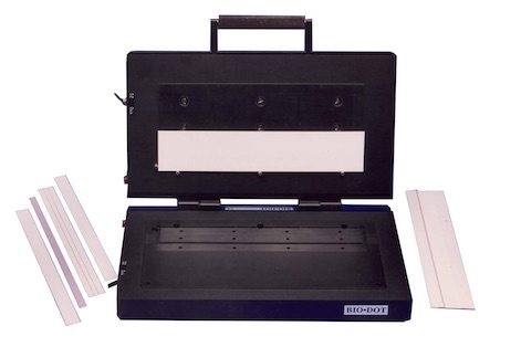
The LM5000 Clamshell assembles a lateral flow assay comprised of multiple materials onto an adhesive backing. It contains top and bottom vacuum nests to hold strip materials in place and is operated manually. Standard and customized nest inserts are available and are easily interchangeable so that multiple assay designs can be laminated.
LM9000

The LM9000 is a continuous laminator which provides continuous lamination of materials onto a plastic support backing with adhesive. This would be used for a assembling rapid diagnostic test strips.
CM5000
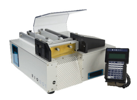
The CM5000 guillotine cutter is also available to provide high quality precision cuts. The widths and cuts can be programmed through a handheld terminal.
DISPENSE PLATFORMS, continued:
The XYZ series systems are a powerful production tool for rapid test development and manufacturing with a larger working deck than the smaller XYZ3060 platform.
XYZ3210
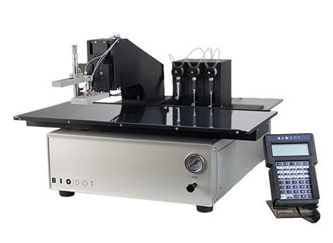
The XYZ3210 is a flexible XYZ motion/dispense system, with a motored Z-axis and fully programmable motion and dispense parameters. The XYZ3210 is designed for simultaneous dispensing or individually dispensing lines and dots. This platform can incorporate 4 AirJet HR, 8 BioJet HR, 8 FrontLine HR, or 8 PolyDrop dispensers. The researcher can choose the type and number of dispensers systems for their application needs.
XYZ3060
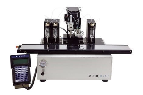
The XYZ3060 system is similar to the XYZ321- system, but with a smaller working deck.
BioDot also provides a variety of automated camera vision solutions in order to increase reproducibility in manufacturing as well as reliability in part inspection. Automated inspection process can lead to a higher quality of product and increased yield. All of these LFA dispensing systems to create your own design and applications can be seen on BioDot’s web site.: https://www.biodot.com/lateral-flow/
Continue to next article: Manufacturing of Lateral Flow Assays (Part 6)
BioDot provides a number of reagent dispensing platforms that are designed for a combination of R&D and production activities. In both cases, the same dispensing technology is used which allows for R&D validated processes to be efficiently transferred to the manufacturing environment.
IMPREGNATION:
Bulk impregnation of sheets in baths or in-line with the RR120 is available. The RR120 has an impregnation module for dipping web materials followed by drying. However, neither of these options are recommended due to the basic non-homgenous nature of these materials which leads to the variations of dried reagent concentrations. This problem is eliminated by the use of the previously described dispensing reagent systems (BioJet HR, AirJet Ultra, AirJet HR, etc). For continuous dispensing, a tandem pump configuration is used for both the FrontLine and BioJet dispensing. BioDot has international patents (US, China, Europe) for the BioJet HR™, BioJet Ultra™, AirJet, as well as the used of solenoid and aerosol dispensed heads combined with the use of syringe pumps to achieve quantitative dispensing. BioDot’s additional patents include the use of the tandem pump configuration, which eliminates delays and line distortions due to syringe refilling cycles.
DISPENSE PLATFORMS:
ZX1010 Platform
The ZX1010 is a flexible dispensing system with a programmable X-axis, manually adjusted Y-axis positioning, and a pneumatic Z-axis. Dispensed volumes are independently programmed via the terminal.
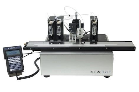
The ZX1010 platform can be configured with a maximum of 8 dispensers (max 4 AirJets). You can choose the type and number of dispensers to meet your application needs and includes the choices of the µAirJet, AirJet HR, BioJet HR, Frontline HR, and PolyDrop dispensers.
Continue to next article: Manufacturing of Lateral Flow Assays (Part 5)
DISPENSING AND IMPREGNATION PRODUCTS, continued:
AirJet HR™
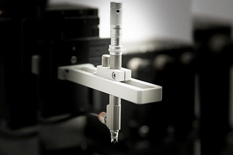
The AirJet HR™ is a nanoliter aerosol dispenser. This BioDot system uses pressurized air to atomize fluid passing through the dispensing nozzle for non-contact, quantitative aerosol dispensing. This creates a quantitative spray format, in which a dot or line can be quantitatively generated on a continuous basis. BioDot’s proprietary technology couples the dispensing nozzle with a high resolution syringe pump to meter exact amounts of reagents. This process produces a precise and easy to use method for dispensing micoliter quantities of fluids.
BioDot’s proprietary technology couples the dispensing nozzle with a high resolution syringe pump to meter exact amounts of reagents. This proces produces a precise and easy to use method for dispensing micoliter quantities of fluids.
µAirJet™
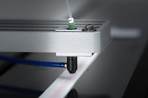
The µAirJet™ is another aerosol type dispenser which uses pressurized air to atomize fluid passing through the nozzle. This nozzle is too, coupled with a syringe pump which creates a spray format. The straighforward design makes µAirJet easy to use, clean, and maintain.
Continue to next article: Manufacturing of Lateral Flow Assays (Part 4)
Order ZosterGent®
Call us toll-free at 888-626-2047 to place an order.
Where to find us
2326 Wisteria Drive, Suite 220
Snellville, GA 30078
United States
Toll-free: 888-626-2047
Main (toll): 678-585-4039
Fax: 678-585-4076
Didn’t find what you need?
Please complete our CONTACT FORM and let us know of your special request!
Don’t miss a thing!
Subscribe to our RSS Feed to be notified when new content is added to the Viro Research website.
Viro Perspectives RSS Feed
