INTRODUCTION:
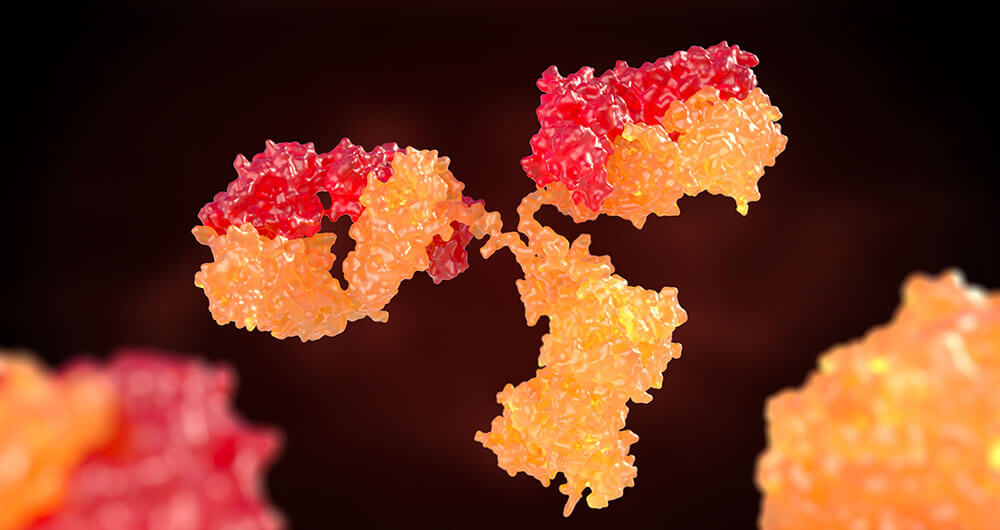
Numerous labs are developing antibody assays to detect COVID-19. There are presently (as of May 1, 2020) four FDA approved assays for detecting IgG/IgM and three for detecting IgG only. Four of these assays use a lateral flow assay (LFA) architecture. All of the assays use the viral S1 glycoprotein as the antibody target. This short note will describe a typical LFA for COVID-19.
DISCUSSION:
A rapid point-of-care assay that detects either/or both IgG/IgM is critical for detecting spread on the infection through the population. A recent infection (<7days) is usually seen with the production of IgM, while older infections (>8 days since infection) detect the generation of IgG. Li et al. (2020) developed an assay that detects both IgG and IgM, detecting antibodies to the SARS-CoV-2 S1 spike protein. They purified the recombinant S1 antigen (MK201027) by protein A affinity chromatography and size-exclusion chromatography. They based the design of the S1 antigen on the published SARS-CoV-2 sequence (MK201027). Antibodies obtained from Sigma include bovine serum albumin (BSA), goat anti-human IgG and IgM antibodies, rabbit IgG, and goat anti-rabbit IgG antibodies. Shanghai KinBio Inc. provided 40Nm gold nanoparticle (AuNP) colloids, NC membrane, and plastic pad, and Whatman provided the glass fiber conjugate (GFC). Sigma produced the PBS. Hunan CDC, China supplied inactivated COVID-19 serum and negative serum samples of patients.
To prepare the AuNP conjugate, they added SARS-CoV-2 recombinant protein dissolved in PBS (1mg/ml) to the mixture of 1ml AuNP colloid (40nm in diameter, OD=1) and 0.1ml of borate buffer (0.1M, pH 8.5). After incubation for 30 minutes at room temperature, the mixture was centrifuged at 10,000 rpm at 4oC for 20 minutes. Next, and 1ml of BSA in PBS was added to the AuNP conjugate to be re-suspended after discarding the supernatant. They repeated the centrifugation and suspension twice, and the final suspension was in PBS. The AuNP-rabbit IgG conjugates were prepared/purified by the same procedure. The main body of the test strip consists of five parts, including plastic backing, sample pad, conjugate pad, absorbent pad, and NC membrane. Each component of the strip is pretreated as follows: the NC membrane was attached to a plastic backing layer for cutting/handling. Researchers immobilized the anti-human-IgM, anti-human-IgG, and anti-rabbit-IgG at test M, G, and control line C. Then, they sprayed conjugate pad with a mixture of AuNP-COVID-19 recombinant antigen conjugate and AuNP-rabbit-IgG. Sample pad was pretreated with BSA (3%, w/v) and Tween-20 (0.5% w/v) before use. To run the assay (at room temperature), researchers pipetted 20 ul whole blood sample (or 10 ul of serum/plasma samples) into the sample port, followed by adding 2-3 drops (70-100ul) of dilution buffer (10mM PBS) to drive capillary action along the strip. The test takes approximately 15 minutes to complete. If only the C line shows red, the sample is negative. Either M or G line or both lines turning red indicates the presence of anti-SARS-CoV-2-IgM or IgG, or both if IgG and IgM are in the specimen.
CONCLUSION:
Of the 397 blood samples (vein blood) from SARS-CoV-2 infected patients, 352 tested positive, for a sensitivity of 88.66%. Twelve of the blood samples from the 128 non-infected patients were positive, for a specificity of 90.63%. Also, 256 out of 397 (64.48%) were positive for both IgG and IgM. Patient finger stick blood was tested and showed that all positive/negative results matched with 100% consistency between vein and finger stick blood, indicating the use of this assay as a point-of-care test using fingerstick blood.
By David Kilpatrick, PhD and Abbas Vafai, PhD
Li, Z., Yi, Y., Luo, X., Xiong, N., Liu, Y., Li, S., Sun, R., Wang, Y., Hu, B., Chen, W., Zhang, Y., Wang, J., Huang, B., Lin, Y., Yang, J., Cai, W., Wang, X., Cheng, J., Chen, Z., Sun, K., Pan, W., Zhan, Z., Chen, L., & Zhang, Y. (2020). Development and clinical application of a rapid IgM‐IgG combined antibody test for SARS-CoV-2 infection diagnosis.
Journal of Medical Virology. https://doi.org/10.1002/jmv.25727
